Connectomics - the practice of mapping every connection in a brain - has improved three orders of magnitude in the last decade, faster than Moore’s law. Milestones keep being passed: the fruit fly brain has been fully reconstructed, a cubic millimeter of mouse cortex is being traced, and experts like Moritz Helmstaedter expect the entire mouse brain to possibly be complete “in the next decade.”
This progress is giving neuroscientists permission to speak seriously about the field’s holy grail: a whole human connectome - a complete wiring diagram of every neuron in a single human brain. At a recent connectomics workshop, organizers noted that naively extrapolating scaling trends “suggest a synapse-resolution whole human brain connectome is technically feasible by 2049”.
But scaling alone understates the pace: recent breakthroughs in sample preparation, imaging, and data processing - among them expansion microscopy, which physically swells tissue several-fold to make visualizing it easier - mean “a far more aggressive timeline is now within reach…decoding the human brain’s circuitry within the next ten years.”
There are many reasons to want this realized as soon as possible. A complete map of the human brain would be an invaluable reference for basic neuroscience, anchoring everything from cell-type taxonomies to models of memory circuitry. Clinical research would benefit too, as most psychiatric and neurodegenerative diseases are, at some level, disorders of connectivity, and comparing connectomes across individuals could surface disease-specific wiring signatures we can currently only infer.
The case also extends beyond basic science and medicine into more esoteric fields. Researchers worried about ensuring increasingly powerful AI respects human values would love to study the wiring diagram of the only system we know that (at least sometimes) produces behavior aligned with them. And those hoping to extend their lives via mind uploading need human brain mapping to become reliable and routine if the method is to give them any chance of escaping death.
A vast and unusually varied set of people, then, will be disappointed to learn that human connectomics faces a problem not hitherto encountered in mapping the brains of other animals: bad-quality brains.
Mapping a brain requires taking a sensitive, living organ and converting it into a manipulable, static object without losing any of its fine details in the process. Of all the body’s organs, brains make this especially hard, as they are exquisitely sensitive to loss of blood flow and shifts in pH, and begin breaking down within minutes if conditions aren’t tightly controlled. Further complicating things, they also have the consistency of soft tofu, liable to tear under their own weight when handled.
These obstacles can be overcome when the brain in question is collected under ideal conditions. The fruit fly connectome was obtained from a fly that was anesthetised and dropped into cold saline; its brain was then rapidly dissected from its head capsule and immersed in preservative chemicals (termed fixatives). Fly brains are small enough - under a millimeter across - that fixative diffuses throughout the entire tissue within minutes, locking its molecular structure in place before any meaningful decay can begin. It also firms the tissue as it goes, transforming the soft, fragile brain into a rubbery solid that can be easily handled in subsequent steps.
Mouse brains are too big for this trick to work. By the time the fixative has diffused from the surface to the deep structures, the interior may already be degrading. Fortunately, their size also makes a more elegant solution available: rather than diffusing fixative inward from the outside, it can be delivered through the brain’s own blood vessels. In a procedure called transcardiac perfusion, an anesthetised mouse has its circulatory system flushed with fixative injected directly into the heart, which carries the chemicals into every capillary in the brain simultaneously. The result is a near-instantaneous transformation of a living organ into a static snapshot - fixed from the inside out and firmed throughout, ready for the next steps in connectomics analysis.
But if a brain isn’t preserved under these near-ideal conditions, connectomics techniques of the foreseeable future have little hope of mapping it.
To understand why, it helps to know what mapping actually involves. After fixation, the brain is sliced into tens of thousands of ultra-thin sections, each imaged under a powerful microscope at a resolution fine enough to resolve all individual cell membranes - what’s known as the tissue’s ultrastructure. Software then traces each neuron through the resulting stack of images, following its membrane from one section to the next to reconstruct its full three-dimensional shape (this process resembles the ‘flood fill’ tool in image-editing software).
What makes this so difficult to achieve is that neurons are extraordinarily long, thin, and densely packed. A single cortical neuron can stretch for centimeters while being less than a thousandth of a millimeter wide, and it must be traced through every single image without error. Mistakes can silently merge two different neurons into one, or split one cell into two phantom fragments. Across an entire brain, these errors compound rapidly: even a 0.1% per-section error rate gives you only a 37% chance of tracing a neuron through a thousand sections cleanly, and essentially zero chance over ten thousand.1
It is troubling, then, that in mice, a delay of as little as ten minutes between loss of normal physiological conditions and the start of fixation can begin to produce exactly this kind of damage - broken membranes scattered throughout the tissue, along with other ultrastructural artefacts that further confound automated tracing.
The brains of larger animals might be somewhat more forgiving. One recent study found largely intact ultrastructure in pig brains when fixative perfusion was delayed by fourteen minutes after circulation ceased, though whether neurons remain traceable across long distances under those conditions has not yet been assessed.
But in any case, the window cannot currently be stretched much further. After roughly fifteen minutes without circulation, reperfusing the brain becomes increasingly impossible - a phenomenon known as ‘no-reflow’, long familiar to stroke clinicians as the reason reopening a blocked artery doesn’t always restore blood flow2. The mechanism is not fully understood, but the leading suspects are swelling of the cells lining the capillaries, contraction of the capillaries themselves, and clumping of blood cells inside the vessels - any of which can physically block fluid from getting through. So even if undamaged neurons are present, once too much time has passed, we don’t currently have a way to quickly deliver fixative to them.
Unfortunately, in the world of human brain banking for research purposes, time-to-fixation after circulation stops is typically measured in hours or days, not minutes. After a patient’s heart stops, the brain is sitting in a warm, oxygen-starved skull through pronouncement of death, transport of the body to the autopsy location, and the start of the autopsy itself - a sequence that usually takes a few hours at a minimum. Admittedly, there are a few research centers running rapid autopsy programs which aim to halt decay within an hour, but they are rare, and even they may not be fast enough for sufficient preservation quality.
To make matters worse, many of these human brains donated to science will have already suffered significant damage in the hours and days before their donor’s heart stopped. A patient in the end stages of cancer or pneumonia doesn’t typically just suddenly die, but instead suffers an ‘agonal phase’ of cascading organ failure, increased blood acidity, and reduced oxygen flow to the brain. By the time their heart gives out and the person officially dies, much damage to their brain has already been done.
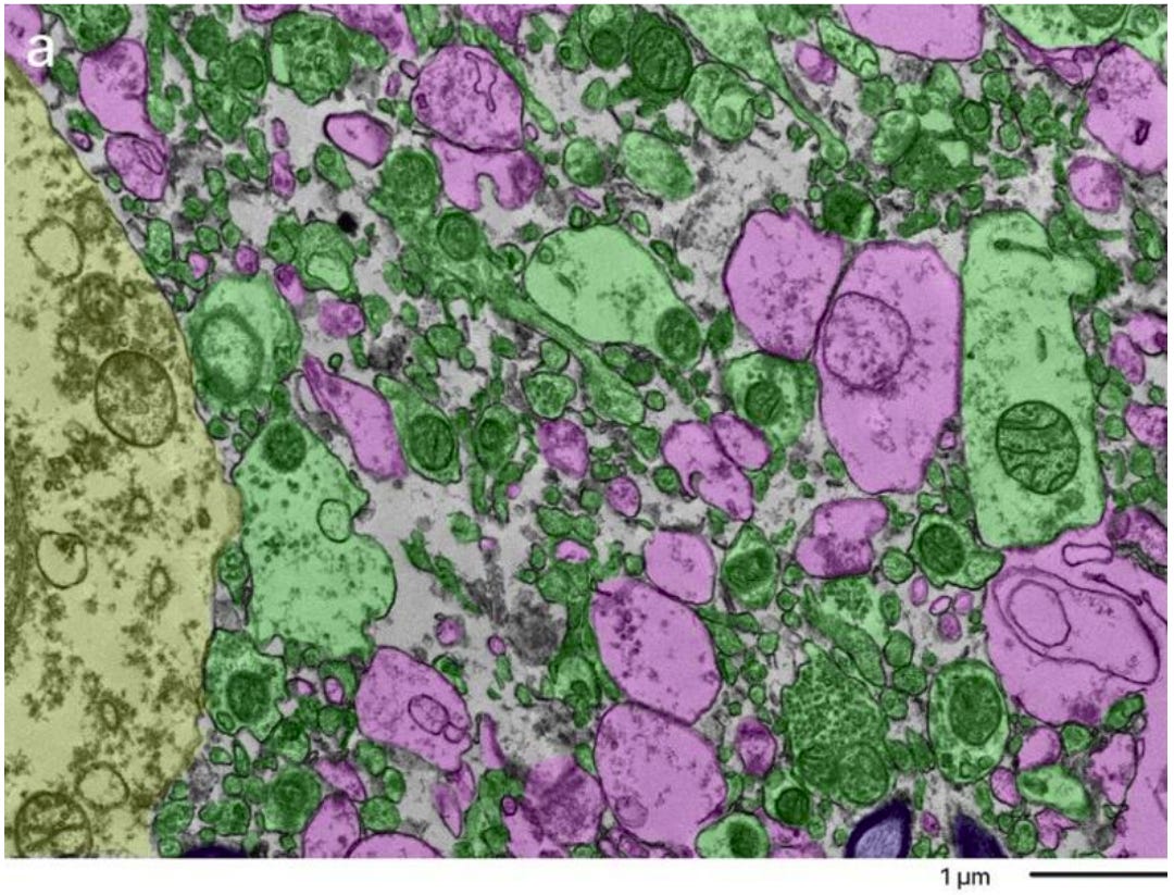
This isn’t to say that fixation under these conditions is useless. Perfusion through the brain’s blood vessels still allows for some flow, gross brain morphology is preserved, and a great deal of cellular structure survives - enough to support traditional neuropathology. But the compromises are visible: as the delay lengthens, the definition of some structures becomes ambiguous, microscopic holes appear, and differences in brain stiffness and blood clearance become apparent even when merely handling the tissue.
For small, carefully chosen volumes, banked tissue can still yield useful data - synaptic ultrastructure has been recovered from human cortex with postmortem intervals of several hours. But that is small-volume nanoarchitecture, not dense, long-range connectomic tracing. Across the distances that matter for following a complete neuron - hundreds of microns to tens of millimeters - banked human tissue is very likely to be riddled with the membrane breaks that make automated tracing impossible.3
What you would actually need then, to have any hope of producing a connectomic-grade human brain, seems close to an impossible set of conditions: a neurologically intact person who dies quickly and predictably, without an agonal phase, in a setting where perfusion fixation could begin within minutes of their heart stopping. To put it more bluntly: you’d need to know in advance when and where someone was going to die, and to have an operating theater standing by.
Conveniently for connectomics research, two organizations are actually already trying to make this happen. Nectome and Sparks Brain Preservation both offer preservation services to terminally ill individuals who hope that preserving their brain will allow them to eventually be revived - by uploading or other means - once technology sufficiently advances. Whatever one thinks of the prospects or ethics of that endeavor, the pursuit of it is pushing both groups to develop techniques for human brain preservation that no one else has yet had reason to develop.
Nectome has been optimising a surgical procedure designed to restore perfusion within minutes of a patient being declared legally dead. To avoid the agonal-phase problem, Nectome takes only patients who are also undergoing medical assistance in dying (MAiD), where particular medication protocols will swiftly lead to cardiac arrest. Their recent preprint reports that the procedure produces electron-microscopy-grade ultrastructure in pig brains (though they’ve not yet shown whether this is sufficient for neuron tracing purposes). If anyone produces a connectomically-tractable human brain in the next few years, it will most likely come from their pipeline.
Sparks Brain Preservation is pursuing a broader research agenda: characterizing the ultrastructure quality of tissue currently held in human brain banks, comparing immersion versus perfusion fixation, and investigating strategies for mitigating the problems caused by long postmortem intervals. Even so, they also operate a preservation facility configured for patients choosing medically-assisted dying, motivated by the same connection between controlled death and high-quality preservation.
If either organization succeeds, the connectomics community will be a major secondary beneficiary. Possibly some of their clients would be willing to consent in advance to having their brains mapped - for those hoping to be uploaded, that is, in the long run, the entire point. But either way, the pipeline itself would become available for ordinary research donors. There are already cases of people undergoing medical assistance in dying being willing to have their final moments studied and their brains subsequently donated. Pairing that willingness with an optimised procedure and a perfusion-ready facility completes the recipe for a connectomics-grade human brain donation.
If we’re to have any hope of mapping the human brain by the end of the century - let alone the more ambitious target of within ten years - then we’ll need an organization like Nectome or Sparks to provide a well-preserved brain sooner rather than later.
It would be complacent for the connectomics community to assume that either party will survive on their own long enough to deliver. Both are currently dependent on futurist-minded investors and a small, slow-growing base of customers with the unusual combination of optimism about eventual revival, the resources to pay for preservation, and access to a jurisdiction that permits assisted-dying. Both are operating and expanding, but this is not obviously the kind of financial base that comfortably sustains long-term research operations.
There is a clear path to derisking the provision of a connectomics-capable preserved human brain. Interested funders could directly support the development of the relevant procedures through grants or investment. Research organizations could purchase preservations in advance for consenting donors, expanding the organizations’ caseloads beyond their current customer base. And neuroscientists could consider working with either of the preservation providers themselves - or those, such as the Brain Preservation Foundation, already seeking to provide third-party evaluation of preservation quality.
All the other fields that stand to benefit from a complete human connectome - neuroscience, medicine, AI capabilities and alignment - are orders of magnitude better funded than the uploaders currently working to remove this bottleneck. For a rounding error in the BRAIN Initiative’s budget, or using the change under the couch at Anthropic, the connectomics community could ensure that human brains are available whenever imaging, segmentation, and processing capabilities finally scale up to meet them.
If you’re a funder considering supporting this work, or a researcher interested in contributing to the development or evaluation of preservation procedures, please get in touch at ariel@brainpreservation.org